What is Magnetic Resonance Imaging (MRI)?
Magnetic resonance imaging (MRI) is a diagnostic procedure that uses a combination of a large magnet, radiofrequency, and a computer to produce detailed images of organs and structures within the body. MRI does not use ionizing radiation, unlike X-rays or computed tomography (CT scans). An MRI provides clear and detailed pictures of internal organs and tissues.

Doctors use MRI to examine soft tissues such as organs, muscle, cartilage, ligaments, and tendons.
It is also helpful when looking at the brain, abdomen, pelvic region and joints like your knee and shoulder. Most MRI exams do not require any special preparation.
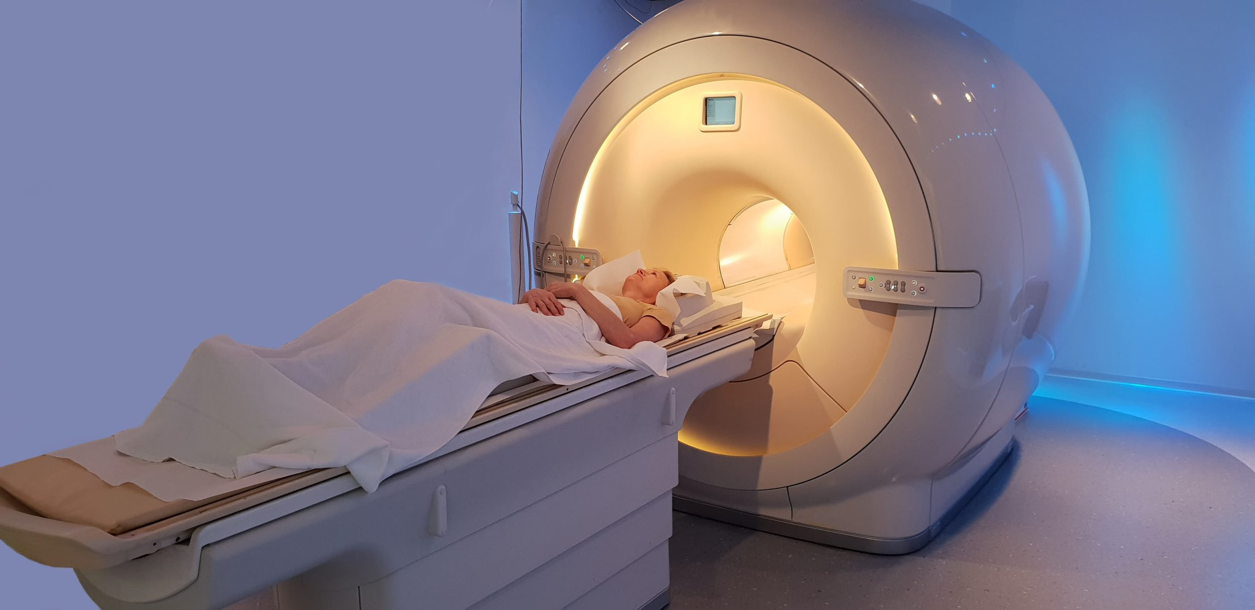
Our NEW Siemens Verio 3T MRI scanner is a state of the art MRI scanner that produces high-quality images especially of the brain, muscles and tendons, bones, joints, and spine. The most important thing for you is the new, open bore 70 cm design which gives you plenty of room to stretch out and feel comfortable. No more tight squeeze!
What People Say About Us!
"I was scheduled for an MRI, and they called me to see if I wanted to come in early! This is fantastic! I came early, checked in very quickly, and as soon as I turned in the paperwork, they took me back to get ready! They were compassionate, understanding, and very professional! The team that actually ran the MRI did a great job getting me comfortable! When it was over, they were very gentle in helping me get up from the table. Well done everyone!"
MRI Services We Offer
- MRI of the brain, neck, spine, chest, abdomen, pelvis, upper and lower extremities
- MR Angiography
- MRCP
- MRI of the Breast
How does an MRI scan work?
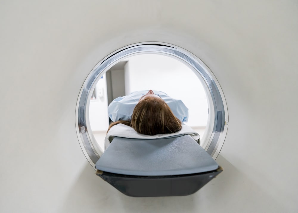
The MRI machine is a large, cylindrical (tube-shaped) machine that creates a strong magnetic field around the patient and sends pulses of radio waves from a scanner. The magnetic field aligns the hydrogen protons in your body along the same vector. The radio waves then knock the particles out of this aligned position.

As the nuclei realign into proper position, the nuclei send out radio signals. These signals are received by a computer that analyzes and converts them into an image of the part of the body being examined. This image appears on a viewing monitor. Cross-sectional views can be obtained to reveal further details. Some MRI machines look like narrow tunnels, while others are more open.
Magnetic resonance imaging may be used instead of computed tomography in situations where organs or soft tissue are being studied because MRI is better at telling the difference between different soft tissues and between normal and abnormal soft tissue.
Reasons for MRI Scan
In orthopedics, an MRI may be used to examine bones, joints, and soft tissues such as cartilage, muscles, and tendons for injuries or the presence of structural abnormalities or certain other conditions such as:
- Tumors
- Inflammatory disease
- Congenital abnormalities
- Osteonecrosis
- Bone marrow disease
- Herniation or degeneration of discs of the spinal cord.
MRI may be used to assess the results of corrective orthopedic procedures. Joint deterioration resulting from arthritis may be monitored by using magnetic resonance imaging. There may be other reasons for your physician to recommend an MRI.
Preparing for an MRI
Prior to your MRI, your physician will explain the procedure to you and offer you the opportunity to ask any questions that you might have about the procedure. If your procedure involves the use of contrast dye, you will be asked to sign a consent form that gives permission to do the procedure. Read the form carefully and ask questions if something is not clear. If IV contrast will be used, patients over the age of 50 years old require a blood test within the past 30 days to measure their Creatinine level.
If you have claustrophobia (fear of enclosed spaces) or anxiety, you may want to ask your physician for a prescription for a mild sedative prior to the scheduled examination. If sedation is used, it is required that you arrange for a relative or friend to drive you home after the exam.
It is important to report any allergies to the radiology technologist. Women should always inform the MRI technologist if there is any possibility that they are pregnant. Many imaging tests are not performed during pregnancy. Also, let your technologist know if you have:
- a pacemaker, aneurysm clips, vascular coils, or filters
- inner ear implants or hearing aids
- unremoved shrapnel or bullet fragments
- insulin or other infusion pumps
- neurostimulator
- permanent dentures
- worked around metal and/or have had fragments in your eye(s)
- surgical shunts
- heart valves
- metal plates, rods, pins or screws
- joint replacements
- surgical staples or wires
Prior to your MRI exam, jewelry and other accessories should be left at home. Because they can interfere with the magnetic field of the MRI unit, metal and electronic objects are not allowed in the exam room. Generally, there is no special restriction on diet or activity prior to an MRI procedure.
Magnetic Resonance Imaging Procedure
MRI may be performed on an outpatient basis or as part of your stay in a hospital. Procedures may vary depending on your condition and your physician’s practices. The entire examination is usually completed within 30 to 45 minutes. If an intravenous contrast material is used, you will feel a pin prick when the needle is inserted into your vein. Some patients may sense a temporary metallic taste in their mouth after the contrast injection.

The technologist begins by positioning you on the MRI exam table, usually lying flat on your back. Straps may be used to help you maintain the correct position and to help you remain still during the exam. A device capable of sending and receiving radio waves will be placed around the area of the body being studied. You will be moved into the magnet of the MRI unit.
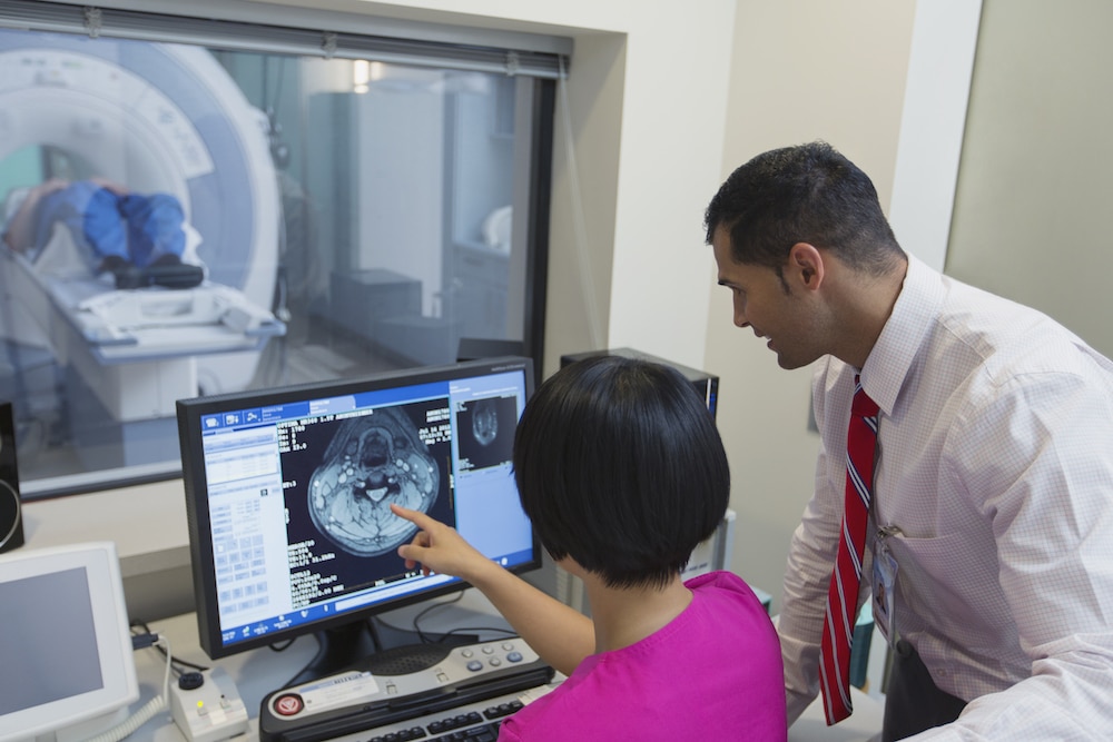
The technologist will be in another room where the scanner controls are located. However, you will be in constant sight of the technologist through a window. Speakers inside the scanner will enable the technologist to communicate with and hear you. You will have a call button so that you can let the technologist know if you have any problems during the procedure. The technologist will be watching you at all times and will be in constant communication.
It is important that you remain perfectly still while the images are being obtained. You will know when images are being recorded because you will hear and feel loud tapping or thumping sounds. Earplugs or headphones are provided to reduce the intensity of the sounds made by the MRI machine. You should notify the technologist if you feel any breathing difficulties, sweating, numbness, or heart palpitations
Once the scan has been completed, the table will slide out of the scanner and you will be assisted off the table. If an IV line was inserted for contrast administration, the line will be removed.
What To Expect After MRI Scan
After the procedure, you should move slowly when getting up from the scanner table to avoid any dizziness or lightheadedness from lying flat for the length of the procedure. If any sedatives were taken for the procedure, you may be required to rest until the sedatives have worn off. You will also need to avoid driving.
If contrast dye is used during your procedure, you may be monitored for a period of time for any side effects or reactions to the contrast dye, such as itching, swelling, rash, or difficulty breathing. If you notice any pain, redness, and/or swelling at the IV site after you return home following your procedure, you should notify your physician as this could indicate an infection or another type of reaction.
Otherwise, there is no special type of care required after a MRI scan. You may resume your usual diet and activities unless your physician advises you differently. A report from your MRI exam will be sent to your doctor within 24 hours.
Online Resources
The content provided here is for informational purposes only, and was not designed to diagnose or treat a health problem or disease, or replace the professional medical advice you receive from your physician. Please consult your physician with any questions or concerns you may have regarding your condition.
This page contains links to other websites with information about this procedure and related health conditions. We hope you find these sites helpful, but please remember we do not control or endorse the information presented on these websites, nor do these sites endorse the information contained here.
- American Academy of Orthopaedic Surgeons
- American Cancer Society
- Arthritis Foundation
- National Cancer Institute (NCI)
- National Institute of Child Health and Human Development
- National Institutes of Health (NIH)
- National Library of Medicine
- National Osteoporosis Foundation
- Osteoporosis and Related Bone Diseases – National Resource Center – NIH
- Radiological Society of North America
