Main Hospital Location
2200 E Show Low Lake Road
Show Low, AZ 85901
Outpatient Pavilion Location
4951 S White Mountain Road
Show Low, AZ 85901
Snowflake Location
1121 South Main Street
Snowflake, AZ 85937
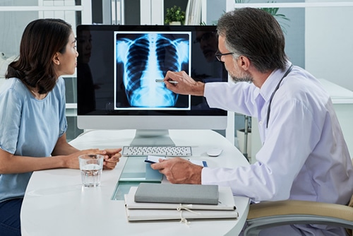
The Diagnostic Imaging Department of Summit Healthcare offers state-of-the-art diagnostic services. Our team includes licensed X-ray technologists, certified CT technologists, mammography technologists and radiologists. Our expert staff complements the comprehensive imaging technology, ensuring excellent patient care and treatment. Our department is on call 24 hours a day, 7 days a week. Providing quality patient care is our primary focus.
Diagnostic imaging employs different technologies to create visual representations of the interior of the body for diagnosis and treatment of disease and other health issues. Although everyone knows the original form of diagnostic imaging — the x-ray — today x-rays have been joined by CT scans, MRIs, PET scans, ultrasound, and other technologies to make a huge impact on diagnostic ability.
Although the x-ray has been used since 1895, it doesn’t distinguish between different soft tissues of similar densities. This made it necessary to perform “exploratory surgery” in many cases so that the surgeon could get an accurate representation of what was going on inside the patient. Today’s advances in imaging technology are revolutionizing diagnosis, and because of it, treatment. Most exploratory surgery is no longer necessary, thanks to these advances.
At Summit Healthcare, we use a variety of forms of diagnostic imaging. We want to provide our patients with the most accurate information possible, and advanced technology aids us in that goal. Here are some of the most common:
- X-Rays
- Magnetic Resonance Imaging (MRI)
- Computer Tomography (CT or CAT)
- Positron Emission Tomography (PET)
- Ultrasound
- Quick diagnostic results
- Less radiation exposure
- Electronic storage of data
- Higher quality images
MRIs use powerful magnets, radio waves, and a computer to make detailed images. Because they don’t use radiation, MRIs can be given more frequently without worry about radiation exposure. MRIs poorly visualize bone, so they are perfect for creating images of the brain and within the spine. MRIs can also be used to check the health of various organs such as the pancreas or liver.
CT scans
PET scans reveal how your tissues and organs are functioning. In PET a chemical compound labeled with a short-lived positron-emitting isotope is injected, swallowed, or inhaled depending on which organ or tissue is being studied. The isotope collects in areas of your body that have higher levels of chemical activity, which can correspond to areas of disease. On the PET scan, these areas show up as bright spots. PET is valuable for detection of cancer and in the evaluation of heart conditions.

Medical ultrasound uses high frequency sound waves that are reflected by tissue to varying degrees. While ultrasound doesn’t provide the anatomical detail of CT or MRI, it can show moving structures in real time. Although associated with imaging of the fetus in pregnant women, ultrasound also is used for imaging of the abdominal organs, heart, breast, muscles, tendons, arteries, and veins.
Nuclear medical imaging procedures often detect a wide variety of conditions such as cancer, heart disease, arthritis and infection. Summit Healthcare was the first hospital in Arizona to acquire a state-of-the-art D SPECT Nuclear Camera.
A mobile Pet Scan is at Summit Healthcare once per week.
- Abscess Drainage
- Arthrogram
- Arteriograms
- Barium Enema
- Bone Biopsy
- Bone Mineral Densitometry / DEXA Scan
- Breast MRI
- CT Scan
- EKG
- Hysterosalpingogram
- Image Guided Biopsy
- Kidney Biopsy
- Liver Biopsy
- Lumbar Puncture
- Lung Biopsy
- Mammography
- MRI Scan
- MRI Ordering Guide
- Myelogram
- Nuclear Medicine Scan
- Paracentesis
- Percutaneous Nephrostomy
- Peripherally Inserted Central Catheter
- PET Scan
- Sonohysterogram
- Stereotactic Guided Breast Biopsy
- Thoracentesis
- Thyroid Biopsy
- Ultrasound
- Ultrasound-Guided Breast Biopsy
- Upper GI Series
- Voiding Cystourethrogram
- Womens Imaging
- X-Ray
Any time there is a question of what is happening inside the body, diagnostic imaging is an effective way to reveal internal structures that could only otherwise be viewed through exploratory surgery. Diagnostic imaging reveals internal structures hidden by the skin and bones, and it allows doctors to have a database of normal anatomy and physiology that can then be compared with the patient’s results to identify abnormalities.
Some exams require preparation; others do not. Depending on the type of imaging exam you are having, your preparation will be different. Some exams may require fasting for 24 hours prior to the procedure, while with others you can eat and behave normally. We will discuss what you need to do, if anything, to prepare for your exam.
- Bring your health insurance card with you to your appointment
- Please provide our staff with 24 hours notice if you need to cancel or reschedule
- Please leave all jewelry and unnecessary valuables at home
- Unless instructed otherwise, take medications as prescribed
- Do not hesitate to call our facility with any questions that you may have
- Carefully follow all examination prep instructions given
- Bring a designated driver if sedation or support is needed
Once your diagnostic imaging exam has been performed, you’ll have a brief wait while we make sure your images have the necessary clarity and resolution, and are oriented correctly. You’ll have a brief appointment with your technician who will explain how the imaging exam went and what the next steps will be. He or she will tell you how long it will take for your doctor to examine your test and provide his or her diagnosis. Most exams don’t require any recovery, but if you were given a sedative you’ll need to have someone else drive you home.
- 64-slice volume
- D SPECT Nuclear Camera
- Acuson S2000 Ultrasound
- Expertise in mammography, Ultrasound, MRI, CT, PET and Interventional procedures
- Modality Accreditations by TheAmerican College of Radiology
- Technologists with advancedlevel training and certifications
- Dedication to patient care andeducation with advanced state-of-the-art imaging services
- Same day appointments
- Full Service Women’sImaging Center
- The most TechnologicallyAdvanced Equipment
- Fully Digitally
- Latest Software
- Sub Specialty Trained Radiologists
- All Radiologists are BoardCertified and ACR Accredited
- Easily AccessibleRadiologist Contact
- Capability to Access reports and Images remotely (real time)
- STAT reports and wet reads
- Copies of images deliveredupon request
- Evening and weekend availability
"Between myself and my family, we have had urgent care and more. This hospital to us is a 5 star hospital! The reason being is they are always a complete team. From checking into surgery or even emergency care; they all work in unison to provide real compassionate care, every time."- Donna C.
